In his popular how-to guide Half Hours with the Microscope, of 1859, the British surgeon and naturalist Edwin Lankester (1814–1874) extolled the virtues of the microscope as a tool through which anyone could gain knowledge of the physical world. “What eyes would be to the man born blind,” he wrote, “the microscope is to the man who has eyes.”1 Christopher Wren (1632–1723), born almost two centuries before Lankester, would surely have agreed. Though much better known as the architect of St. Paul’s Cathedral in central London, the Royal Hospital in Chelsea, and the Old Royal Naval College in Greenwich, Wren played a now all but forgotten but pivotal role in popularizing microscopy. When his drawings of magnified fleas and lice were shown to Charles II in 1661, the king was so intrigued that he asked Wren and the Royal Society to make such magnifications more widely accessible to the public through their publication.
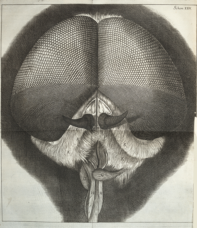
Wren was too busy with architectural projects at the time to take on the task, but he convinced his friend and fellow microscopist Robert Hooke to do so. Over the next four years, Hooke illustrated and wrote about objects and organisms he and Wren were able to observe in astonishing new detail. In 1665 the Royal Society published his treatise under the title Micrographia: or Some Physiological Descriptions of Minute Bodies Made by Magnifying Glasses with Observations and Inquiries Thereupon. This magnificent book, long prized by collectors, details fifty-seven microscopic observations of specimens at various degrees of magnification, starting with the point of a needle, and going on to such disparate subjects as silk, hair, plants, seeds, and a number of insects with which humans were uncomfortably familiar—a fly, a flea, and a louse.2 The publication ended with three short essays in which Hooke moved on to the very largest and most distant subjects he could see by using a telescope: stars and the moon. There had never been a book quite like it. The English diarist Samuel Pepys was so besotted by it, that he famously recorded sitting up until two o’clock in the morning reading the book, declaring it “the most ingenious book that ever I read in my life.”3
Brilliantly illustrated with detailed engravings of his subjects (Fig. 4), Hooke’s book is often considered the starting point for the popularization of microscopy in Western science.4 Certainly, among the members of England’s Royal Society and other wealthy, well-educated people, it was a foundational publication. It took almost two hundred years for microscopes (and telescopes) to become sufficiently affordable to the middle class to make such interesting subjects more widely accessible.5
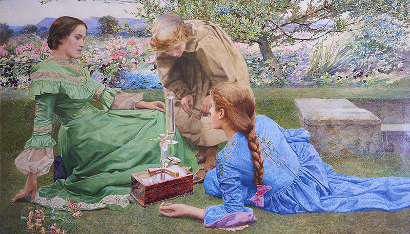
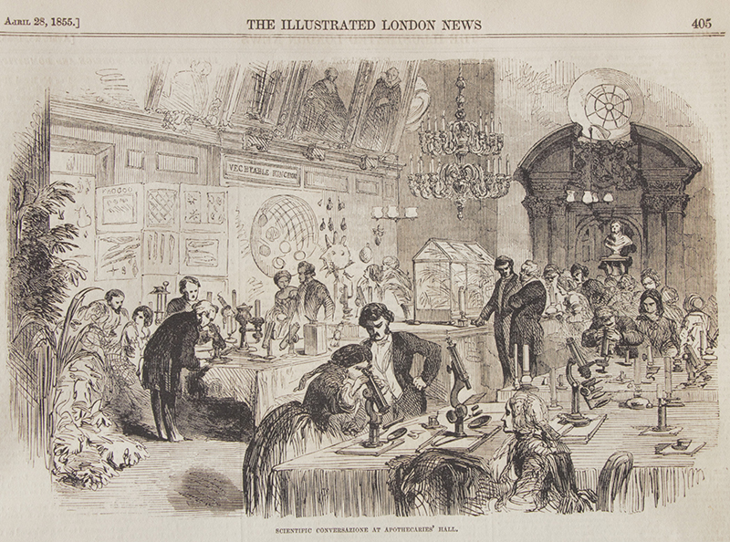
By the middle of the nineteenth century, the magnifying device that Hooke had brought to the public’s attention exploded in popularity, and with it came a new way of looking at the world. “The microscope is rapidly becoming the companion of every intelligent family that can afford its purchase,” wrote Henry Slack in 1861.6 For those with enough leisure and a curiosity about science, a great deal of time was spent with a microscope, often coupled with trips to the seashore searching for shells and seaweeds, or traipsing through fields and forests picking wildflowers, grasses, and ferns for closer scrutiny. Such activities, beautifully conveyed by the English painter Lexden L. Pocock in a watercolor titled The Microscope (Fig. 2) and in a wood engraving depicting a more public “conversazionoe” in 1855 published in the Illustrated London News (Fig. 6), embodied the “rational amusements” so eagerly sought by the Victorian middle class.
A number of private societies and clubs dedicated to microscopy and other scientific pursuits were established throughout Great Britain and in the United States during the mid- to late 1800s. These allowed enthusiasts to gather with likeminded friends and colleagues for lively discussions and to share visual delights. The pooled resources of club members enabled the purchase of high-end microscopes and other apparatus that might have been too expensive for individual members. The frequent meetings of these groups were as much social gatherings as scientific exchanges.7
Of course, microscopes had been accessible to the wealthy for some time through the universities and other academic institutions with which they were associated. Anyone else willing and able to pay the cost of admission in select cities in Britain and Europe could view subjects magnified to previously unimaginable scale at public venues. Beginning at the end of the eighteenth century, solar and oxy-hydrogen microscopes were used to attract—and sometimes shock—audiences with public presentations of otherwise invisible subjects.8 With these instruments, “the monstrous forms [found] in a single drop of water”9 (Fig. 5) and other live subjects like “a flea or a louse [shown] as big as a goose or a jackass”10 were projected onto large screens and interpreted by experts. One viewer described the stunning effect of the subjects he saw projected on a fourteen-foot screen: “A few hairs of an infant appeared like tubes two inches in diameter,” he wrote. “The sting[er] of a bee was a monstrous barbed weapon, four feet long. The lancets of the horsefly were sabers about two feet in length. . . . Some of the worms found in stagnant ditches, the natural size of which is that of a thread, appeared like the largest-sized boa constrictor.”11
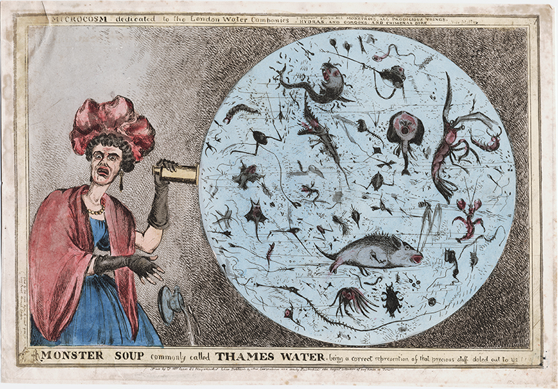
After viewing a projected microscope show in London in March 1833, the German Prince Pückler-Muskau (1785–1871) reacted with horror to what was revealed to be living in drinking water: “What [the microscope] shows,” he wrote, “is really enough to drive a man of lively imagination mad. Nothing can be more horrible,—no more frightful devilish figures could possibly be invented,— than the hideous, disgusting water animalculae (invisible to the naked eye, or even to [magnifying] glasses of inferior power,) which we daily swallow. They looked like damned souls darting about their filthy pool with the rapidity of lightening, while every motion and gesture seemed to bespeak deadly hate, horrid torture, warfare, and death.”12
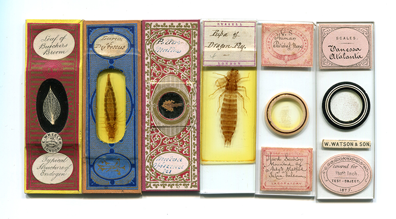
When science-oriented amateurs were able to view microscopic subjects in the quiet of their own homes, they tended to focus on less disturbing subjects, preferring slides of inert specimens that were chosen as much for their beauty as for what they could tell their viewers about the natural world. Among the most popular subjects for microscopic enjoyment were diatoms, the single- celled, aquatic organisms whose silica shells come in a wide variety of intricate, exquisitely beautiful shapes and patterns. Although they lie at the base of many of nature’s most important food chains and can be found in both fresh and salt water around the world, until the widespread use of microscopes, diatoms had been largely unnoticed by all but aquatic specialists because they are essentially invisible to the naked eye. In the nineteenth century they became sources of great wonder and the centerpieces for many a family conversation and collection.
Unlike snowflakes, whose crystalline structure they sometimes resemble, diatoms are stable and their shells long-lasting. As with other algae, they are classified by genus and species. Their beauty, stability, and identifiable place in the hierarchy of scientific classification appealed to amateur naturalists seeking to understand the organization of nature’s many living organisms. And so microscopic slides holding diatoms became widely popular. They were sometimes prepared by the microscopists themselves, but more often traded with friends or purchased from commercial slide sellers (Fig. 7).
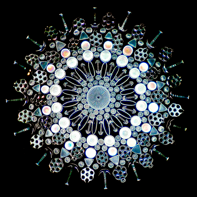
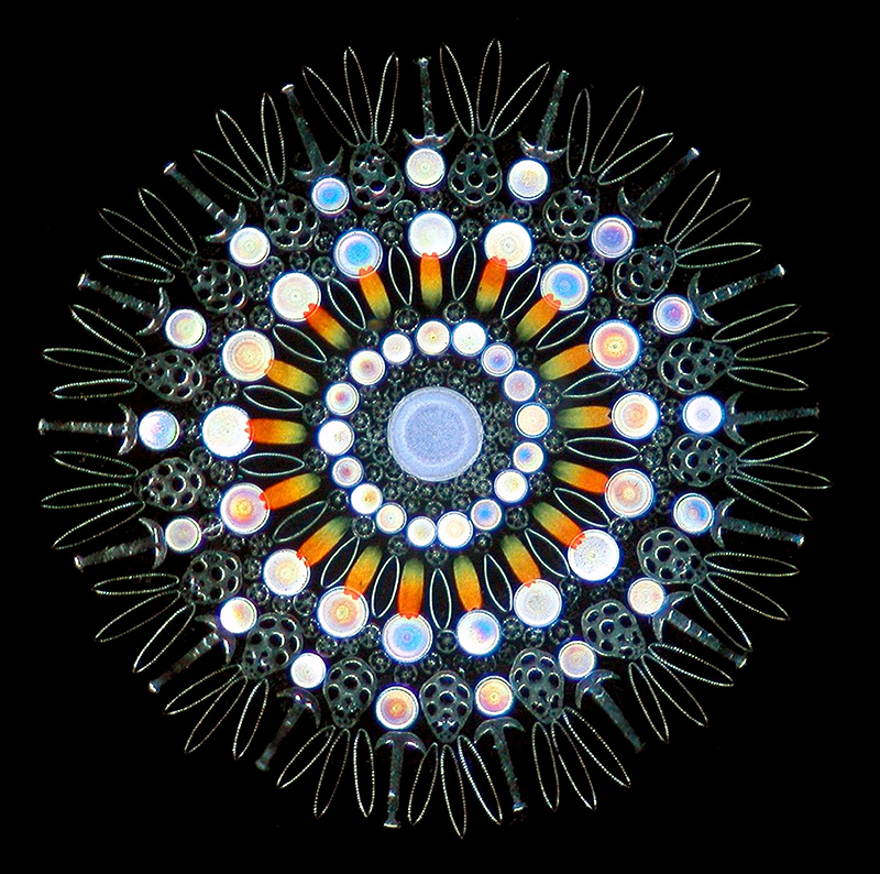
Complementing the inherently beautiful shapes of the individual diatom crystals, some slide makers arranged their subjects in geometric patterns. Two of the best such makers, Johann Diedrich Möller and Eduard Thum were German, though their microscope slides were eagerly acquired by microscopists from around the world. Möller had originally planned to pursue a career in the fine arts and went to art school to perfect his skills, but he became infatuated with microscopy along the way. He ultimately decided to establish a business creating microscope slides to which he could apply his artistic talents. He organized some of his subjects by species, and some by geographic location, but many of his slides were eclectic gatherings of different species, from different places, arranged in geometric patterns for the sheer beauty of their display (Fig. 8). Making such slides required not only sensitive aesthetic judgment but took an enormous amount of time and manual dexterity. Möller clearly had all three.
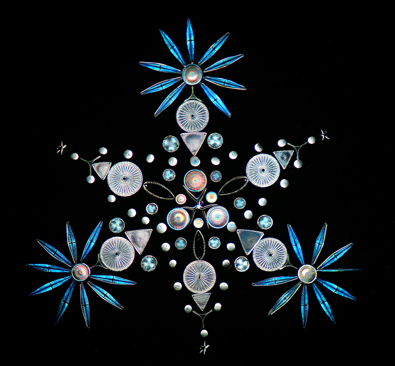
Thum sometimes made slides of thin-sectioned rocks, chemical crystals, and other materials, but his favorite subjects were diatoms. These he arranged by species, presenting perfect frustules (the siliceous part of a diatom cell wall) of each in different positions to permit the study of their structures. Like Möller, Thum also prepared “salon” style slides using diatoms, butterfly scales, and other minute objects. These he arranged into star shapes and other geometric patterns (Figs. 1, 9). Some of his groupings were given the shapes of flowers, birds, and other subjects in which beauty, not scientific classification, was the intended subject.
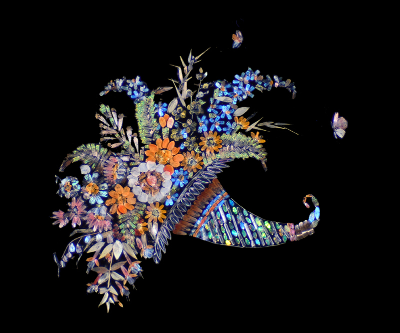
The man who surpassed all others in creating artistic compositions on a microscopic scale was Henry Harold Dalton. The son of a prominent physician and born and raised in Suffolk, England, Dalton eventually moved to the Continent and settled in France. By 1875, when he was praised for his talents in the Monthly Microscopical Journal, he was supplying individual “micromosaics” to collectors around the world.13 In the US, one of his admirers was William H. Walmsley (1830–1905), a founder of the American Society of Microscopists (now the American Microscopical Society) and an active member of the Academy of Natural Sciences of Philadelphia. At an evening meeting of the microscopical section of the academy, Harvey shared with his colleagues “a resplendent slide of butterflyscales arranged by Mr. Harold Dalton, of Havre, representing birds of brilliant plumage, around an iridescent vase.”14 We don’t know exactly which of Dalton’s creations Harvey displayed, but since many of his surviving slides feature compositions matching this general description (Figs. 10–12), we can understand why it was considered worthy of note in the academy’s records.
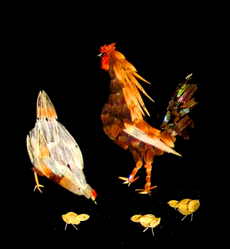
To make these amazing mounts, Dalton stripped the scales from butterfly wings and arranged them by size, shape, and color on a miniature palette. Next, working with a special microscope designed for the purpose, he transferred the scales to a glass slide using a single boar bristle. He would then position the scales using a thin glass tube through which he blew tiny puffs of air. As each scale arrived in the place he wanted it, he would press it to the glass. There, its natural oils would secure it in place until the entire composition was complete and he could protect it with a glass cover slide. According to an 1886 article in the American journal Popular Science News, Dalton “was a fast worker, and by laboring almost incessantly [on this task], he could finish [a single slide] . . . in the course of a week or ten days.”15 A single slide could have as many as a thousand individual scales.
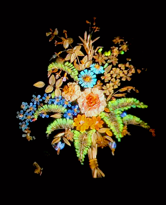
The Popular Science reporter described one of Dalton’s decorative slides as having “eighty two distinct flowers of various shades and colors, and each as perfect as it would be possible for an artist to represent it on canvas.” Describing this as “one of the wonderful achievements of the century,” he went on to say that Dalton’s slides were “as highly prized by microscopists throughout the world as a rare painting by a celebrated master is prized.”16 At the time, the magazine reported, there were “not more than fifty Dalton slides in this country [the US],” which could “scarcely be purchased for love or money.”17 That number remains about the same today, as does their coveted status among microscope slide collectors.
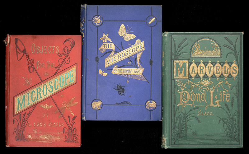
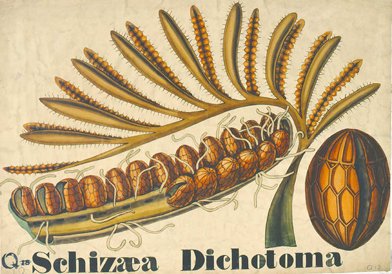
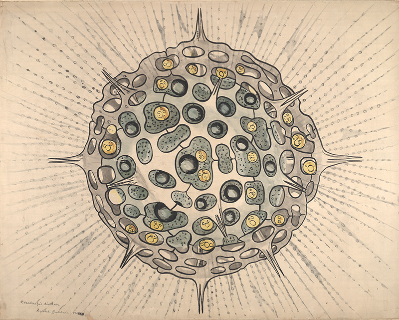
For those who didn’t have microscopes of their own and were more interested in the details of nature than the artistic compositions microscope slide makers could create, there were illustrated books (Fig. 13), magazines, and, for classroom use, large posters and teaching charts with microscopic subjects. In the 1830s and ’40s, the British botanist John Stevens Henslow, Charles Darwin’s friend and mentor, produced a series of spectacular teaching charts of previously unseen botanical subjects using a microscope (Fig. 14). Forty years later, the American naturalist-educator Joseph Leidy, sometimes called the father of parasitology because of his pioneering work in that field, created hundreds of large teaching charts of his own, thus enabling his students to see the organisms or parts of organisms on which he focused his lectures, from the back of a large lecture hall (Fig. 15). The designs of the Henslow and Leidy charts provide good examples of the seamless intersection of science and art that was prevalent in the nineteenth century. They are as beautiful as they are informative and embody the (then) new way of seeing the world through the magical and transformative lens of a microscope.
Many of people who created microscope slides, wrote books about microscopy, and painted the wall charts that made minute organisms big enough for all to see, modestly claimed they were merely helping to advance the public’s understanding of science and drawing attention to God’s creations. This may have been true, but by saying so, they undervalued their own remarkable talents and contributions. The originality and artistic skill that made their works of art so dazzling to today’s eyes have earned for their makers our attention and praise. Although it is something they would never have sought, recognition of their work for its own sake is well-deserved.
1 Edwin Lankester, Half-Hours with the Microscope (London 1859). See also John R. Dolan, “From the Popularization of Microscopy in the Victorian Age: A Lesson for today’s ‘Outreach,’” Protist, vol. 170, no. 3 (July 2019), p. 7, and online at sciencedirect.com. 2 A few of Hooke’s illustrations appear to have been plagiarized from De Nivis usu Medico Observationes Variae by Thomas Bartholin (1616–1680), published four years earlier, in 1661. Hooke’s microscopic illustrations were reissued in 1745 and continued to be reproduced in microscopy books throughout the nineteenth century. See Margaret ‘Espinasse, Robert Hooke (London: William Heinemann, 1956), p. 59. 3 Entry for January 21, 1664/65, in Samuel Pepys’s diary, available at pepysdiary.com. 4 There were other earlier books that contained illustrations of microscopic subjects, including Olaus Magnus’s Historia de Gentibus Septentrionalibus of 1555, which reproduced the earliest known picture of a snowflake, but Hooke’s book had the greatest impact. See Brian Ford, Images of Science: A History of Scientific Illustration (Oxford: Oxford University Press, 1993). 5 Lynn Barber, The Heyday of Natural History (Garden City, NY: Doubleday, 1980), p. 121. In an advertisement in The Lens, the quarterly journal of microscopy published by the State Microscopical Society of Illinois, in 1872 (vol. 1, no. 1), the James Queen microscope company, with salesrooms in Philadelphia and New York, was selling “instruments of every grade, from $3.00 to $1300 each” including the “unequalled” Immersion Objective made by R. and J. Beck of London for $50. In subsequent issues of the journal, where microscope slides are offered at prices ranging from 50 cents to $1, the editor warns against buying too inexpensive a microscope, stating that “No decent efficient instrument can be obtained under a cost of almost $50, and from that prices run up easily to $2000” (The Lens, vol. 1, no. 4 [November 1872], p. 247). 6 Henry J. Slack, Marvels of Pond Life or A Year’s Microscopic Recreations (London 1861), p. 3. 7 Some of these clubs have continued to meet up to the present day. A typical example is today’s Leidy Microscopical Society, originally formed in 1858 as the Microscopial Society of Philadelphia, which retains its historic collection of slides and microscopes and continues to meet regularly. 8 The oxy-hydrogen microscope, which projected microscopic subjects onto a screen using limelight, magnifying lenses, and a magic lantern, entertained audiences from 1825 through the first decades of cinema. See Meegan Kennedy, “Throes and struggles . . . witnessed with painful distinctness”: The oxy-hydrogen microscope, performing science, and the projection of the moving image,” Victorian Studies, vol. 62, no. 1 (Autumn 2019), pp. 85–118. 9 Literary Gazette, July 19, 1928, p. 463, quoted in Richard Altick, The Shows of London (Cambridge MA: Belknap Press of Harvard University Press, 1978), p. 370. 10 Quoted in S. Bradbury, The Microscope Past and Present (Oxford: Pergamon, 1968), p. 117. 11 Quoted in Altick, The Shows of London, p. 370, citing Mirror, vol. 21 (1833), pp. 138–139, in the Enthoven Collection, Theatre Museum, London. 12 [Hermann Ludwig Heinrich von Pückler-Muskau], Tour in England, Ireland, and France, in the Years 1826, 1827, 1828, and 1829 . . . by a German Prince (Philadelphia, 1833), p. 172, quoted in Altick, The Shows of London, p. 370. 13 Brian Stevenson, “Henry ‘Harold’ Dalton, 1836–1912,” micro scopist.net, citing Augustus de Souza Guimaraens, “English and foreign preparers of microscopic specimens,” Monthly Microscopical Journal, vol. 14 (October 1875), pp. 209–210. 14 Philadelphia Medical Times, vol. 5 (June 5, 1875), p. 574, quoting the proceedings of the meeting of April 5, 1875, of the Biological and Microscopical Section of the Academy of Natural Sciences. I am indebted to Brian Stevenson and Howard Lynk for their extensive research on many of the leading microscopists of the nineteenth century, and to David Wilson, founding director of the Museum of Jurassic Technology, which has one of the largest collections of Dalton slides, for this information. I recommend Howard Lynk’s “A Cabinet of Curiosities: A Selection of Antique Microscope Slides from the Victorian Era c. 1830s–1900” at victorianmicroscopeslides.com/slides.htm as the single best online source for information on this subject. 15 “Painting Microscopic Slides,” Popular Science News, vol. 20, no. 8 (August 1886), p. 108. 16 Ibid. 17 Ibid.